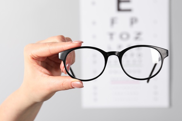
Is Retinal Photography Painful?

Early signs of many eye diseases may show up on the retina, or the back of the eye. retinal photography is a way of evaluating the back of the eye through color, high-resolution, digital photographs on which such signs may show up more easily.
Taking the images does not hurt at all. However, it does require the eyes to be dilated with special eye drops. After your eyes have been dilated, you will be sensitive to light for several hours. Because the photography takes place in a dark room, this is not bothersome until your appointment is over and you leave the office. You may need to wear dark glasses to shield your eyes from the light and have someone else drive you home. Eventually, the drugs that cause your eyes to dilate wear off and your pupils start reacting to light as usual.
Why is retinal photography performed?
As digital imaging of the retina becomes more advanced, efficient, and affordable, it may be performed more often. As of now, however, it is usually not performed as a part of routine eye exams. However, if your optometrist identifies symptoms of potential eye disease as part of routine screening, retinal photography may be ordered to get a clearer, more detailed image of the back of the eye and to confirm or rule out a diagnosis.
Digital imaging of the retina shows a number of important structures in the eye:
- Blood vessels that provide circulation to the eye
- The optic disk, where the optic nerve connects to the eye
- The macula, which allows the eye to focus and produce clear images
The following are eye conditions that retinal photography can help identify.
Glaucoma
Glaucoma occurs when the pressure inside the eye increases. This compresses the blood vessels in the eye and puts pressure on the optic nerve, resulting in vision loss.
Diabetic retinopathy
Diabetes can damage blood vessels in the eye, causing them to leak or swell. It can also cause new blood vessels to form. Early detection of diabetic retinopathy can prevent blindness through interventional treatment.
Macular degeneration
Most often associated with age, macular degeneration causes a loss of central vision because of changes to the blood vessels in the macula. With wet macular degeneration, which is the more severe type, the changes are due to the development of abnormal blood vessels in the macula. With dry macular degeneration, the existing blood vessels go through changes that cause them to become brittle and thin.
If you are diagnosed with an eye disease such as these, you may continue to have retinal photography performed at routine intervals to monitor your condition and further assess your condition. Even if you do not have an eye disease, photographing the retina offers the additional benefit of preserving a permanent record of changes in your eye over time.
Conclusion
Retinal photography can be performed to track changes in the eye and to diagnose ocular diseases. While the procedure itself is painless, it does require pupil dilation, which can cause discomfort for several hours thereafter until the medication wears off.
If you are experiencing any symptoms of possible eye disease or it has been a while since your last examination, contact our office to schedule an appointment.
Check out what others are saying about our services on Yelp: Retinal Photography in Dallas, TX.
Recent Posts
A contact lens exam addresses different questions than a standard eye exam. It evaluates the cornea, lids, and tear film, then matches lens materials and designs to vision needs and eye health. Lenses sit directly on the eye, so small details such as curvature, diameter, and oxygen flow influence comfort, clarity, and safety. An optometrist…
Standard vision tests, or eye exams, help ensure accurate prescriptions for glasses and support long-term eye health for people who wear corrective lenses. Even small prescription changes can affect headaches, screen comfort, and night driving clarity. Eye exams also screen for conditions that may not present with early symptoms, making routine testing an important habit.…
Pediatric eye care is the key to clear vision and confident learning that starts in childhood. Unfortunately, even minor vision problems can negatively affect reading, coordination, and classroom focus. Many parents come to realize how common it is for children to pass quick screenings yet struggle with focusing, eye strain, or fatigue during schoolwork. A…
The selection of eyewear feels significantly easier when the styles and craftsmanship of Versace eyewear enters the conversation, since bold lines and iconic detailing create an instantly recognizable look. Versace represents the intersection of Italian fashion and Greek mythos, blending confident glamour with symbolism through its signature Medusa logo. The brand is widely associated with…